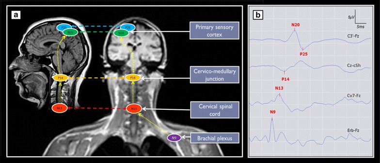
Fig. 2.

Schematic representation of median nerve’s somatosensory evoked potentials (SSEP) responses localizations on brain MRI (a) and typical examples of their normal wave forms as well as recording electrodes montages (b). SSEP elicited by electric stimulation (15 mA) of median nerve at the wrist: N9, N13 and P14, respectively, the brachial plexus, cervical spinal cord, and cervico-medullary (subcortical) responses. N20 and P25 are responses of the primary sensory cortex. N9–N13 inter-peak latencies (IPL) represent a proximal peripheral nerve conduction time, and P14–N20 IPL the intracranial conduction time (ICCT). Recording and reference electrodes were placed at Cv7 (7th cervical vertebra)—Fz: for the N13 cervical spinal cord response and Cz-cSh (contralateral shoulder): for the subcortical far-field potential: P14. The cortical components, N20 and P25 were recorded at the contralateral C3′ or C4′ positions (2 cm behind C3 or C4) according to the international 10–20 system. Two sets of 500 sweeps were averaged
