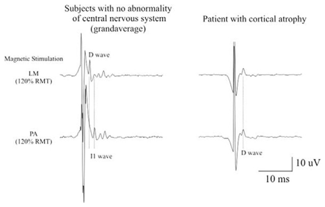
Figure 2.

Epidural activity recorded in a patient with cerebral cortex atrophy. Descending volleys evoked by latoro-medial magnetic stimulation and posterior-anteriormagnetic stimulation at 120% resting motor threshold (RMT) in five patients with no abnormality of the central nervous system and at the maximum stimulator output in one chronic alcoholic patient. The grand averages of epidural volleys recorded in patients with no abnormality of central nervous system are shown on the left and the averages of epidural volleys (of 10 sweeps) recorded in the patient with cerebral cortex atrophy are shown on the right. The latencies of the D and I1 waves evoked by LM and PA magnetic stimulation are indicated by vertical dotted lines. In control subjects, LM stimulation evokes a large D wave followed by 5 I waves; PA stimulation evokes only I waves. In the patient with cerebral cortex atrophy the output evoked by LM and PA magnetic stimulation is similar. Both techniques evoke a large D wave and no clear I waves but only two very small and delayed peaks (Di Lazzaro et al., 2004b).
