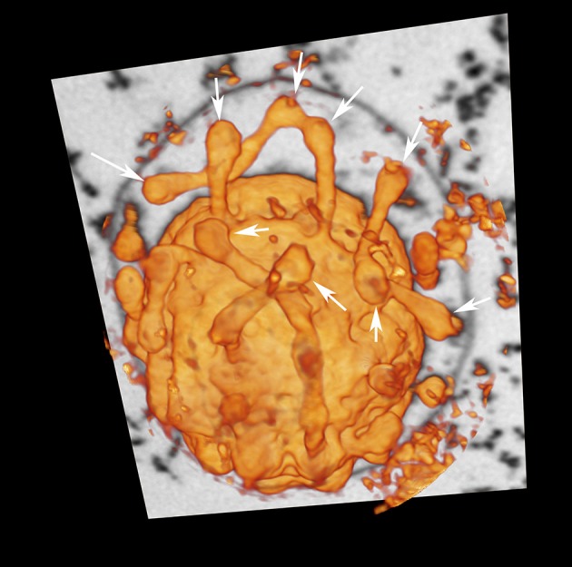
Figure 2.

Cytoplasmic protrusions of I. hospitalis. “Voltex” (volume texture rendering) display of a I. hospitalis cell from the FIB/SEM data showing protrusions from the cytoplasm; arrows point to spherical swellings that might indicate constriction or fusion sites, respectively; additionally one slice of the image stack of the original data is shown, in which the OCM can be seen.
