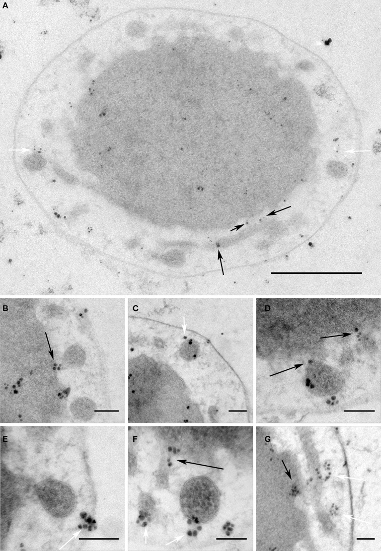
Figure 6.
V4R protein Igni_1332 immunolabeling. Immunolabeling on 50 nm sections against Igni_1332 showing (A) a whole I. hospitalis cell and (B–G) additional examples from different I. hospitalis cells; black arrows point to examples for localizations in the IM and the membrane of protrusions at putative fusing sites; white arrows point to labeling associated with the matrix of filaments and/or tethers in the IMC; bar 0.5 μm (A); 100 nm (B–G)

