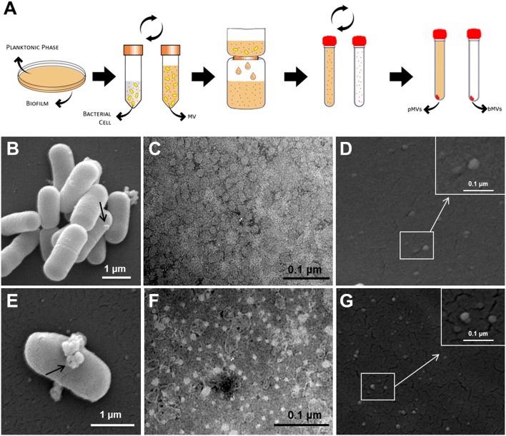
FIGURE 2.
Lactobacillus reuteri DSM 17938 membrane vesicles isolation from planktonic and biofilm phenotypes. pMVs and bMVs isolation procedure (A); SEM image of a biofilm sample containing L. reuteri cells, which generate extracellular vesicles (arrow) (B); SEM image of a planktonic cell producing multiple vesicles (arrow) (E); Negative staining analysis of bMVs (C) and pMVs (F); vesicles released from L. reuteri biofilm cells (D) and planktonic cells (G) detected by SEM. Magnification of MVs (Square insert). Representative images of six independent experiments.

