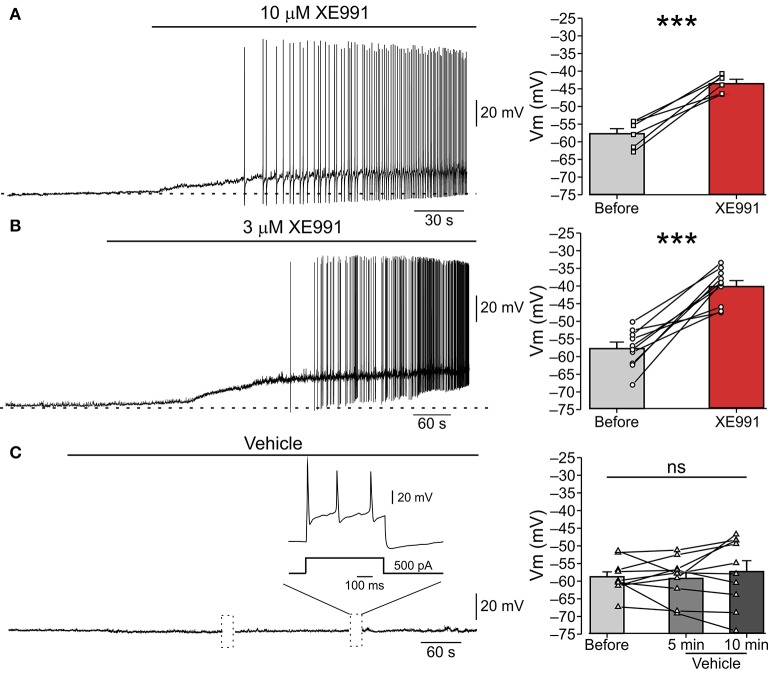
Figure 1.
XE991 leads to spontaneous firing of nociceptor-like DRG neurons. (A) Left, Typical responses of nociceptor-like small (25 μm) DRG neurons to focal puff application of 10 μM XE991 (representative of 5 out of 6 experiments). Current clamp perforated-patch recordings performed from acutely dissociated DRG neurons perfused with extracellular solution. Note, membrane depolarization and onset of spike discharges shortly after application of XE991. Dashed line indicate resting potentials before drug application (−58 mV). Right, mean ± SEM (bar graphs) and individual neurons' changes in membrane potential before (gray) and after application of XE991 (red, measured before generation of first action potential). ***p < 0.001, paired Student t-test, n = 6. Note that XE991-induced depolarization in all recorded neurons. (B) Same as in (A), but 3 μM XE991 was puff-applied on the neurons. Dashed line indicate resting potential before drug application (−59 mV). Left, representative of 10/11 neurons. ***p < 0.001, paired Student t-test, n = 11. Note that 3 μM XE991 induced depolarization in all recorded neurons. (C) Control experiment. 10 min long puff application of vehicle onto nociceptor-like DRG neuron. Note, no spontaneous depolarization developed during this period, yet the cell fired normally upon injection of depolarizing current pulses (inset; representative of 9 experiments). Dotted boxes show time breaks in free-run recordings when current protocols where applied. Right, same as in (A,B), but measured 5 min and 10 min after puff application of vehicle; ns—not significant, one-way ANOVA, n = 9.

