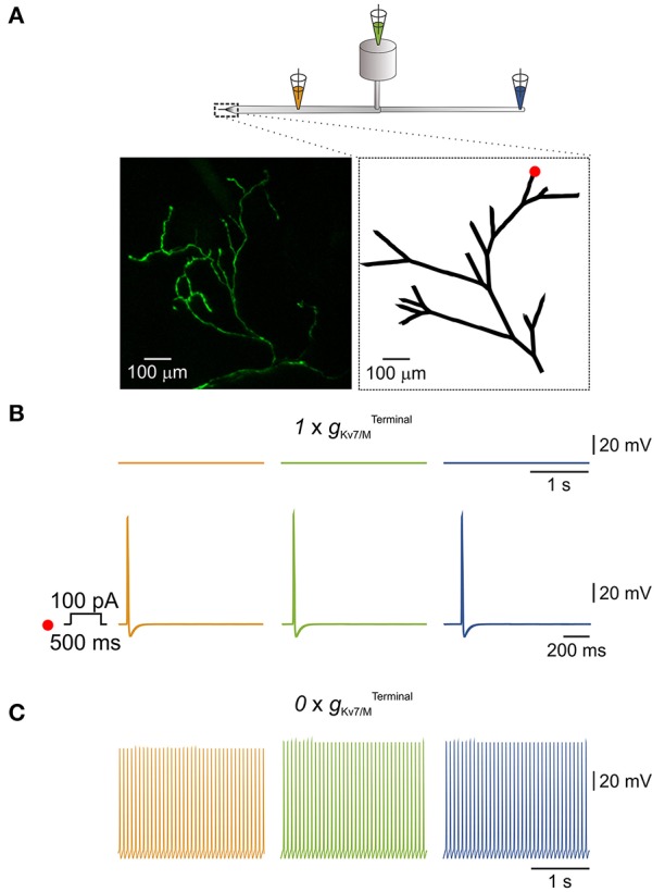
Figure 4.

IM in the terminals is sufficient to prevent spontaneous activation of a simulated multi-compartment nociceptive neuron. (A) Scheme depicting the general structure of the multi-compartment model. Dotted box outlines the nociceptive terminal, enlarged in the right inset below. Orange, green, and blue pipettes indicate the sites from which the changes in membrane potentials were recorded and correspond to: peripheral axon (orange), soma (green), and central axon (blue). Insets, Left, in-vivo multiphoton imaging of nociceptive peripheral terminals and distal axons in the mouse hind paw expressing GFP (z-stack; see Methods). Right, geometry of the terminal tree based on the rendering of the image shown in left which was used in the NEURON environment to simulate nociceptive peripheral terminals and distal axons. The red dot indicates the stimulus loci. (B) Upper traces, recordings from peripheral axon (orange), soma (green), and central axon (blue) when terminal Kv7 conductance () was intact (, see Methods). Note, no spontaneous action potential firing, yet application of 100 pA depolarizing current for 500 ms at the distal terminal branch (indicated as a red dot in A, inset, right) lead to generation of a single action potential which propagated along the neuron. (C) Annulling of () lead to generation of spontaneous action potential firing at the distal axons (orange) which fully propagated along the neuron (green and blue).
