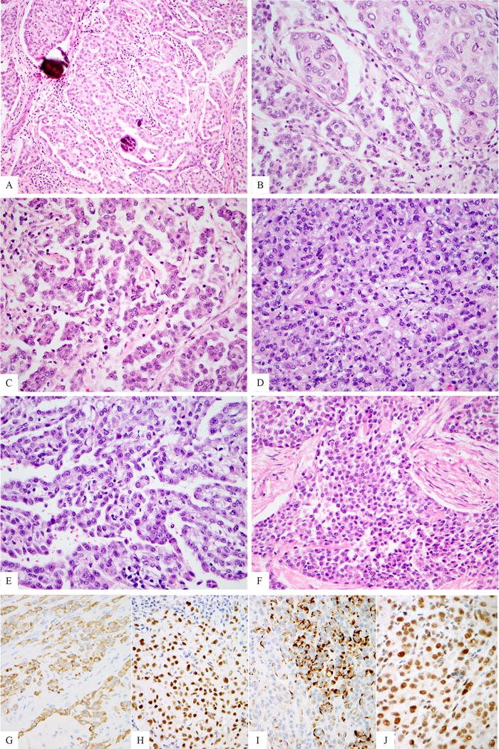
Figure 1. Histologic features of EWSR1-ATF1 fusion positive mesotheliomas.

Medium power view of peritoneal index case (MM1) exhibiting focal psammoma bodies (A). The tumor displayed a conventional epithelioid morphology with focal papillary architecture, as well as abundant eosinophilic cytoplasm and open chromatin (B, C). MM2 showed a predominantly solid growth (D), with only focal papillary architecture (E). MM3 exhibited a predominant round cell phenotype with scant cytoplasm (F). All cases were immunohistochemically positive for AE1/AE3 (G, MM2), WT1 (H, MM2) and focal desmin expression was observed in a single case (I, MM2). Immunohistochemical expression of BAP1 was retained in the 3 fusion positive cases tested (J, MM2).
