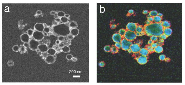
Figure 7.
EFTEM spectrum-image from plastic sections of hybrid nanocomplexes containing heparin, protamine, and ferumoxytol, which are being used for magnetic labeling of stem cells. (a) Fe L2,3 edge image; (b) overlay of Fe L2,3 edge image (red), S L2,3 edge image (blue) and N K edge image (green) shows that the iron-containing ferumoxytol nanoparticles surround a soft core composed of sulfur-containing heparin and nitrogen-containing protamine. The two components in the core co-localize due to association between the negatively charged heparin and the positively charged protamine. From L.H. Bryant et al. [38].

