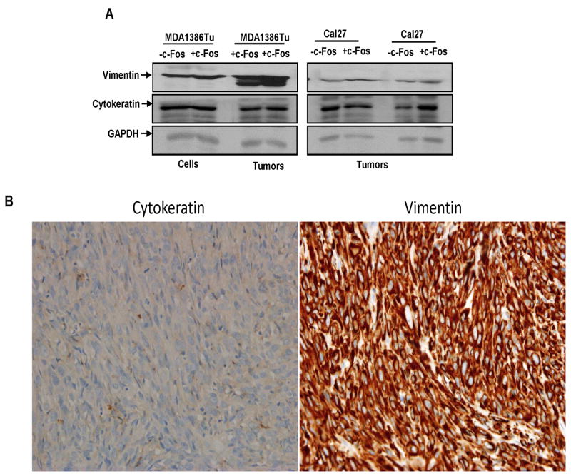
Figure 2.
Increased expression of vimentin from c-Fos overexpressing MDA1386Tu cells. (A) Lysates from Cal27, MDA1386Tu control and c-Fos expressing cells and tumors are subjected to Western blot analysis for vimentin and cytokeratin expression using specific antibodies. Blots are reprobed with an antibody to actin for comparison of protein loading in each lane. (B) Immunohistochemistry images showing expression of vimentin and cytokeratin from MDA1386Tu-c-Fos cells implanted tumors. Images are taken at 40x.

