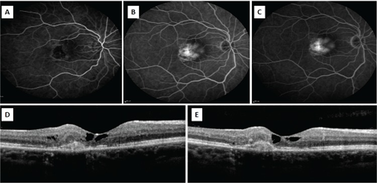
Figure 2. A-C) Fundus fluorescein angiography in a patient with type 2 juxtafoveal telangiectasia shows hyperfluorescence due to subretinal neovascularization (SRNV) beginning in the early phase and increasing in the later phases. D) Optical coherence tomography reveals the internal limiting membrane, foveal atrophy, and SRNV and intraretinal fluid temporal of the fovea. E) Regression of the intraretinal fluid is observed after intravitreal injection.

