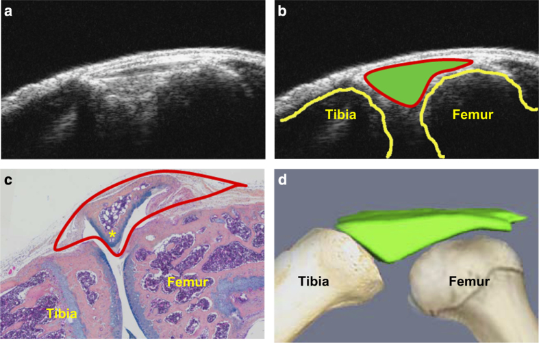
Figure 1.
Orientation of joint space on an ultrasound image. A 3-month-old wild-type C57BL/6 male mouse was used. (a) An ultrasound B-mode image of a mouse knee joint. (b) Outlined joint space (solid green), tibia, and femur on the ultrasound B-mode image. (c) Alcian blue/Orange G (ABOG)-stained knee section with outlined joint space detected by ultrasound. (d) Illustration shows 3D reconstruction of join space derived from a stack of ultrasound B-mode images.

