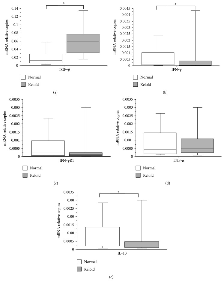
Figure 3.
(a) Number of mRNA relative copies for TGF-β present in biopsies of patients with keloid compared with that of patients in the control group (Mann–Whitney; p < 0.001). (b) Number of mRNA relative copies for IFN-γ present in biopsies of patients with keloid compared with that of patients in the control group (Mann–Whitney; p = 0.009). (c) Number of mRNA relative copies for IFN-γR1 present in biopsies of patients with keloid compared with that of patients in the control group (Mann–Whitney; p = 0.246). (d) Number of mRNA relative copies for TNF-α present in biopsies of patients with keloid compared with that of patients in the control group (Mann–Whitney; p = 0.911). (e) Number of mRNA relative copies for IL-10 present in patients with keloid biopsies compared to that in patients in the control group (Mann–Whitney; p = 0.037). The horizontal line represents the median, the bar percentile of 25% to 75%, and the vertical line the percentile of 10 to 90%. ∗ indicates significant p value.

