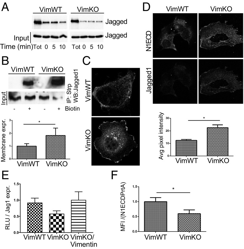
Fig. 3.
Vimentin regulates Jagged recycling and Notch activation. (A) Analysis of Jagged 1 recycling in VimWT and VimKO cells using a biotin cell surface labeling and stripping protocol. (B) Cell surface proteins were labeled with biotin and immunoprecipitated using streptavidin agarose beads. Immunoblotting was performed with an antibody detecting Jagged 1. (C) Representative confocal microscopy images showing Jagged 1 immunofluorescence in VimWT and VimKO cells. (D) Representative confocal microscopy image shows Jagged 1 cell surface localization and N1ECD binding in VimWT and VimKO cells. (Scale bar, 10 µm.) The graph shows quantification of pixel intensity. Values represent means ± SEM. Statistical significance was determined using Student’s t test, P < 0.05. (E) Jagged signal sending potential measured by coculturing VimWT and VimKO cells and VimKO cells reexpressing vimentin with 293HEK cells expressing the Notch 1 receptor (293HEK-FLN1) using a luciferase-based reporter system. Jagged signal sending potential as related to surface levels of Jagged. Values represent means ± SEM. Statistical significance was determined using Student’s t test, P < 0.05. RLU, relative light unit. (F) The ability of VimKO and VimWT to internalize N1ECD–Alexa-488 coupled to protein A agarose beads (N1ECDPrtA). Internalization of N1ECDPrtA in VimKO and VimWT cells as related to surface levels of Jagged. Values represent means ± SEM. Statistical significance was determined using Student’s t test, P < 0.05. MFI, mean fluorescence intensity.

