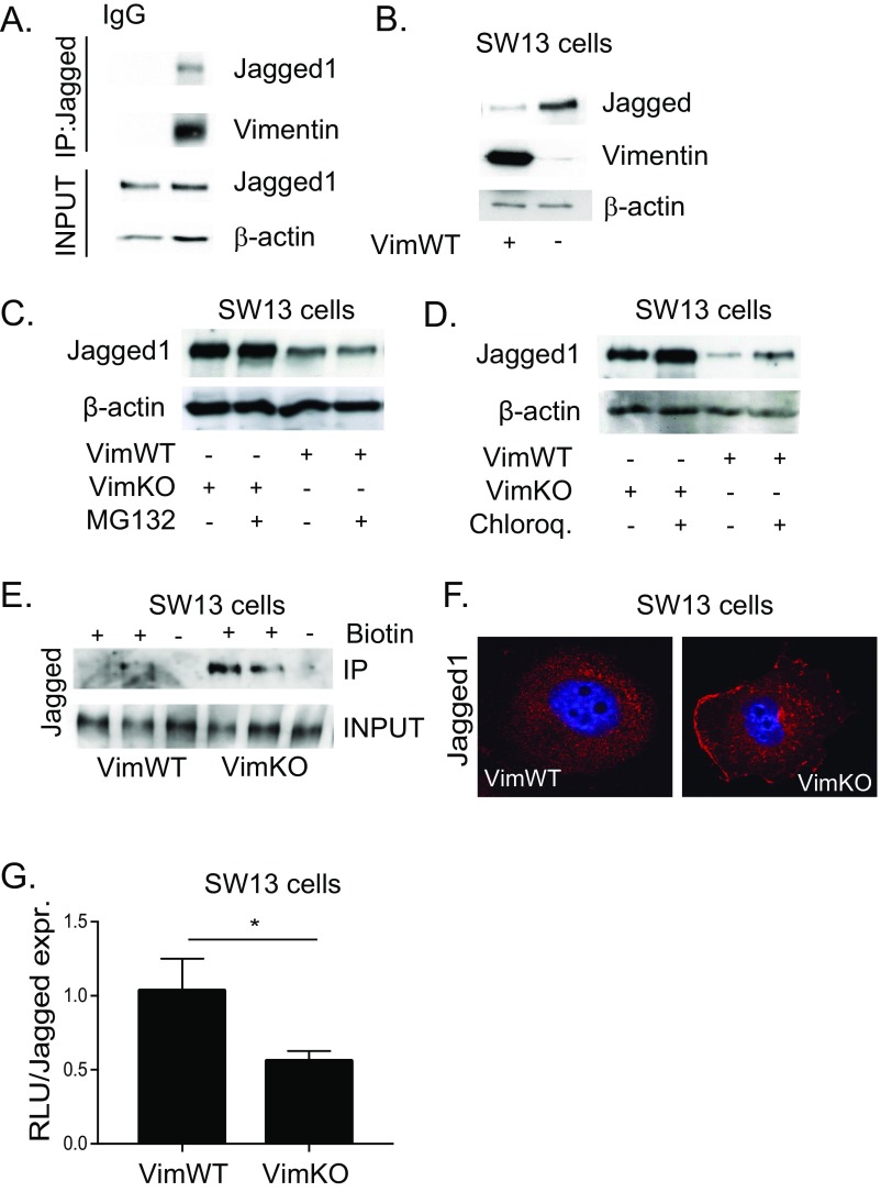
Fig. S2.
Vimentin does not modulate Jagged turnover. (A) Vimentin coimmunoprecipitates with Jagged in 293 HEK cells transfected with vimentin. (B) Representative immunoblot shows Jagged 1 protein levels in vimentin-negative and -positive SW13 cells. (C) Representative images showing Jagged 1 protein expression in vimentin-negative and -positive SW13 cells in the presence of the proteosome inhibitor MG132 (20 µM, 6 h). (D) Representative immunoblot showing Jagged 1 protein expression in vimentin-negative and -positive SW13 cells in the presence of the lysosomal inhibitor chloroquine (20 µM, 16 h). (E) Cell surface proteins in SW13 cells were labeled with biotin and immunoprecipitated using streptavidin agarose beads. Immunoblotting was performed with an antibody detecting Jagged 1. (F) Representative confocal microscopy images showing Jagged 1 immunofluorescence in vimentin-negative and -positive SW13 cells. (G) Jagged signal sending potential measured by coculturing vimentin-negative and -positive SW13 cells with 293HEK cells expressing the Notch 1 receptor (293HEK-FLN1) using a luciferase-based reporter system. Jagged signal sending potential as related to surface levels of Jagged. Values represent means ± SEM. Statistical significance was determined using Student’s t test, P < 0.05. RLU, relative light unit.

