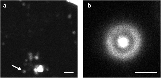
Fig. 4.
Representative confocal microscopic images of HEK293T/17 cells that have been incubated with 1 μM 4CN-Trp*-MpX for ∼10 h, obtained with a scanning step size of 100 nm (A) and 50 nm (B). (Scale bar in A, 10 μm; in B, 2 μm.) Many blebs and other cellular debris can be seen in A, indicating that the HEK cells have died. The higher-resolution image in B corresponds to the bleb indicated by the white arrow in A.

