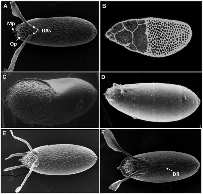
Fig. 1.
Drosophila eggshell, a complex structure derived from an epithelial sheet. (A) Scanning electron microscopy (SEM) image of the eggshell of D. melanogaster. The most prominent features are the two dorsal appendages (DAs), the micropyle (Mp), and the operculum (Op). (B) The egg chamber midway through oogenesis stained with phalloidin. (C) SEM image of the eggshell resulting from uniform activation of DPP signaling in the follicle cells. (D) SEM image of the eggshell resulting from reduced EGFR signaling. (E) SEM image of the eggshell from D. virilis, a species with four dorsal appendages. (F) SEM image of the eggshell from D. willistoni, a species with two dorsal appendages and a dorsal ridge (DR). All SEM images present the dorsal views of the eggshell (anterior to the left).

