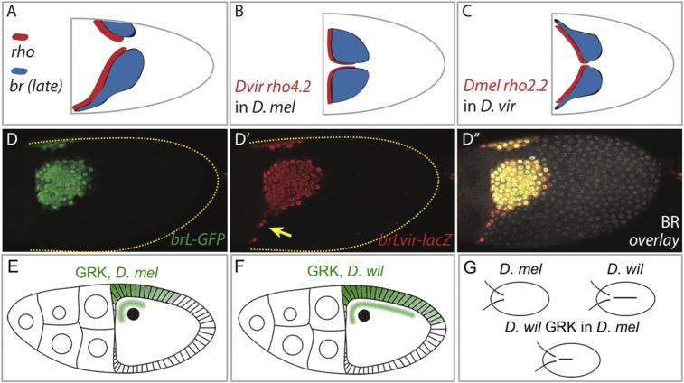
Fig. 4.
Two different mechanisms for the evolution of br patterning: changes in the regulatory region of br and changes in the inductive signals. (A) Schematic representation of the expression domains of br and rho in D. virilis. (B) Summary of the activity of the rho enhancer from D. virilis in D. melanogaster. (C) Summary of the activity of the rho enhancer from D. melanogaster in D. virilis. (D–D′′′) Comparison of the activities of the brL enhancers from D. melanogaster (D; brL-GFP in green) and D. virilis (D′; brLvir-lacZ, red), analyzed in D. melanogaster. (D′′) Merged image including immunostaining for endogenous Br (white nuclear staining) for orientation. The yellow arrow indicates the lateral expansion of D. virilis brL. (E) Schematic of GRK localization in D. melanogaster (lateral view). (F) Schematic of GRK localization in D. willistoni (lateral view). (G) Schematics of eggshells in D. melanogaster, D. willistoni, and D. melanogaster patterned by GRK from D. willistoni.

