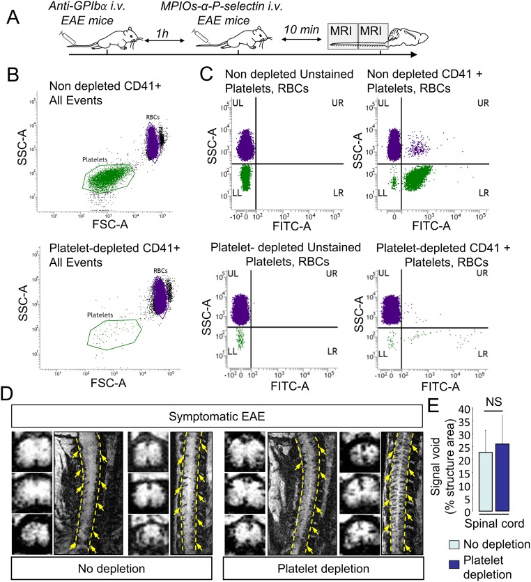
Fig. S2.
MPIOs-αP-selectin–induced hyposignal is not due to binding in platelets. (A) Schematic representation of experimental design. (B) Flow cytometry analyses of anticoagulated whole blood in nondepleted and depleted animals. Forward- and side-scatter density plots show that RBC (purple dots) and platelet (green dots) populations are clearly distinguishable based on their respective light scatter patterns. (C) Box plot analyses of blood samples upon staining with anti-CD41 show platelet-positive events (green dots). (D) Representative high-resolution T2*-weighted images after MPIOs-αP-selectin injection in depleted (Right) and nondepleted (Left) animals. (E) Corresponding signal void quantification (n = 4 per group). NS, not significant.

