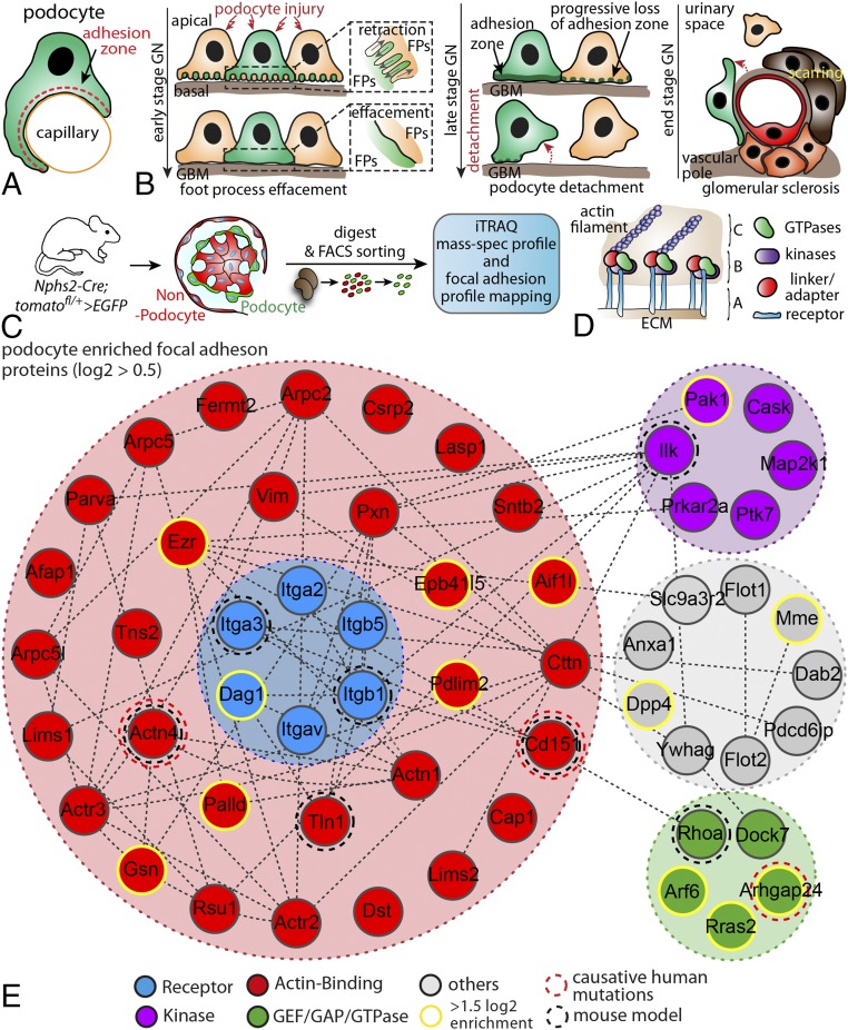
Fig. 1.
Analysis of the podocyte-enriched adhesome identifies EBP41L5. (A) Podocytes reside on the outer surface of glomerular capillaries and attach via their foot processes to the glomerular basement membrane. (B) Schematic depicting different stages of podocyte damage and restructuring of FA complexes (C) Scheme for the generation of hNPHS2Cre*Tom.GFP-reporter mice. Cre expression under the specific hNPHS2 promotor resulted in selective expression of GFP in the podocyte population. Isolated GFP+ cell populations were further processed for iTRAQ-based MS analysis. (D) FAs are composed of different classes of scaffold and signaling proteins such as GTPases, specific kinases, or structural linker molecules (A, integrin receptors; B, adaptor proteins and enzymes; C, actomyosin cytoskeleton). (E) Mapping of the podocyte-enriched FA complex after filtering of primary data sets against the gene ontology term FA. Previously identified proteins involved in human disease or analyzed in rodent models are highlighted by dotted circles. Via clustering resulting from enrichment scores, a subset of proteins were selected as highly enriched (yellow circles; for enrichment scores and selected proteins, see Dataset S1). ECM, extracellular matrix; FA, focal adhesion; FACS, fluorescence-activated cell sorting; GBM, glomerular basement membrane; GN,glomerulonephritis; iTRAQ, isobaric tag for relative and absolute quantitation.

