Fig. 3.
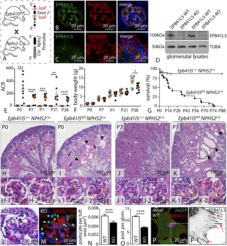
Podocyte-specific knockout of Epb41l5 causes nephrotic syndrome and lethality. (A) Schematic illustrating the generation of a podocyte-specific knockout mouse (B and C) Immunofluorescence staining confirmed that EPB41L5 protein is not detectable in the podocyte compartment in respective knockout mice. (D) Western blot on glomerular lysates from either control or respective knockout animals revealed that EPB41L5 protein is completely abolished. (E and F) Proteinuria measurements demonstrate a drastic increase of proteinuria in Epb41l5 knockout animals beginning at P0 (at least n = 7; Dataset S3), accompanied by decreased body weight gain (at least n = 10 animals per group; SI Appendix, Dataset S3). (G) Kaplan-Meier analysis indicated premature death of Epb41l5 knockout animals (at least n = 15; Dataset S3). (H–K) Histology of wild-type and Epb41l5fl/fl*NPHS2Cre kidney sections revealed proteinaceous casts (black arrows), dilated tubules (black arrowheads), mesangial proliferation (yellow asterisk), and mesangiolysis (yellow arrows). (L–O) PAS and immunofluorescence staining for WT-1 demonstrated detachment of podocytes in Epb41l5 knockout animals. Quantification of WT-1-positive cells in KO animals at 3 wk of age (n = 3 animals for each genotype; Dataset S3). (P) EPB41L5 localized in a typical FA pattern in primary wild-type podocytes (white box indicates zoom-in; red arrows indicate FAs). ACR, albumin to creatinine ratio.

