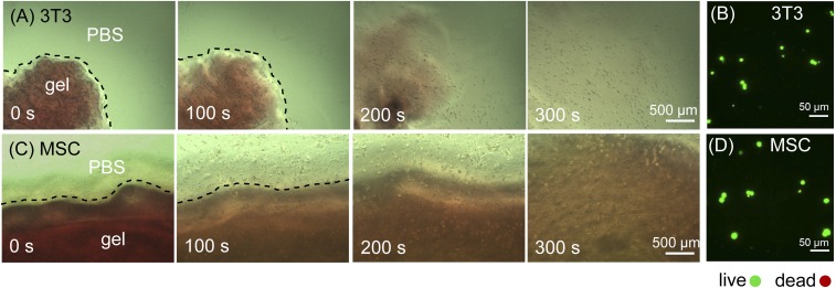
Fig. 4.
Release of encapsulated cells by light-induced gel-sol transition. (A) Mouse 3T3 fibroblasts and (C) hMSCs were encapsulated by CarHC hydrogels (red) and cultured for 24 h. Cell release was initiated by exposing the 3D cell culture to white light (22 klux). Representative bright-field micrographs (0, 100, 200, and 300 s after light exposure) are shown. Dashed lines indicate the gel boundaries. (B and D) Live/dead staining of recovered 3T3 fibroblasts and hMSCs.

