
Figure 2.
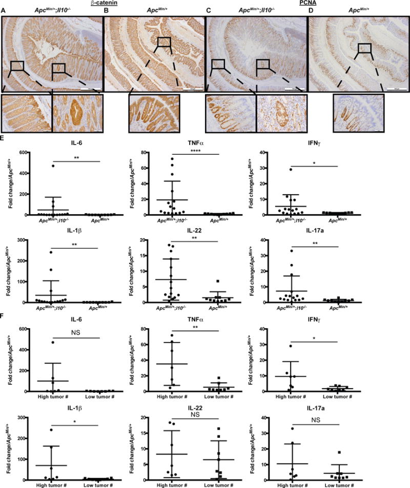
ApcMin/+;Il10−/− mice have increased colon proliferation and inflammation.
A-B) CTNNB1 immunohistochemistry (IHC) from ~16 week old SPF ApcMin/+;Il10−/− (A) and ApcMin/+ (B) colons. C-D) PCNA IHC from SPF ApcMin/+;Il10−/− (C) and ApcMin/+ (D) colons. Higher magnification of both dysplastic and normal regions are shown for SPF ApcMin/+;Il10−/− mice. E) IL-6, TNFα, IFNγ, IL-1β, IL-22 and IL-17a mRNA expression in 16–48 week old SPF ApcMin/+;Il10−/− and ApcMin/+ proximal colon tissue snips with relative fold expression compared to ApcMin/+ mice. F) IL-6, TNFα, IFNγ, IL-1β, IL-22 and IL-17a mRNA expression in 16–48 week old SPF ApcMin/+;Il10−/− stratified by tumor number (high: > 2 tumors or low: ≤ 2 tumors) with relative fold expression compared to ApcMin/+ mice. Data are expressed as mean +/− SD. Two-tailed Mann-Whitney statistical analysis: ****p< 0.0001, ***p< 0.001, **p< 0.01, *p< 0.05, NS: not significant.