Figure 2.
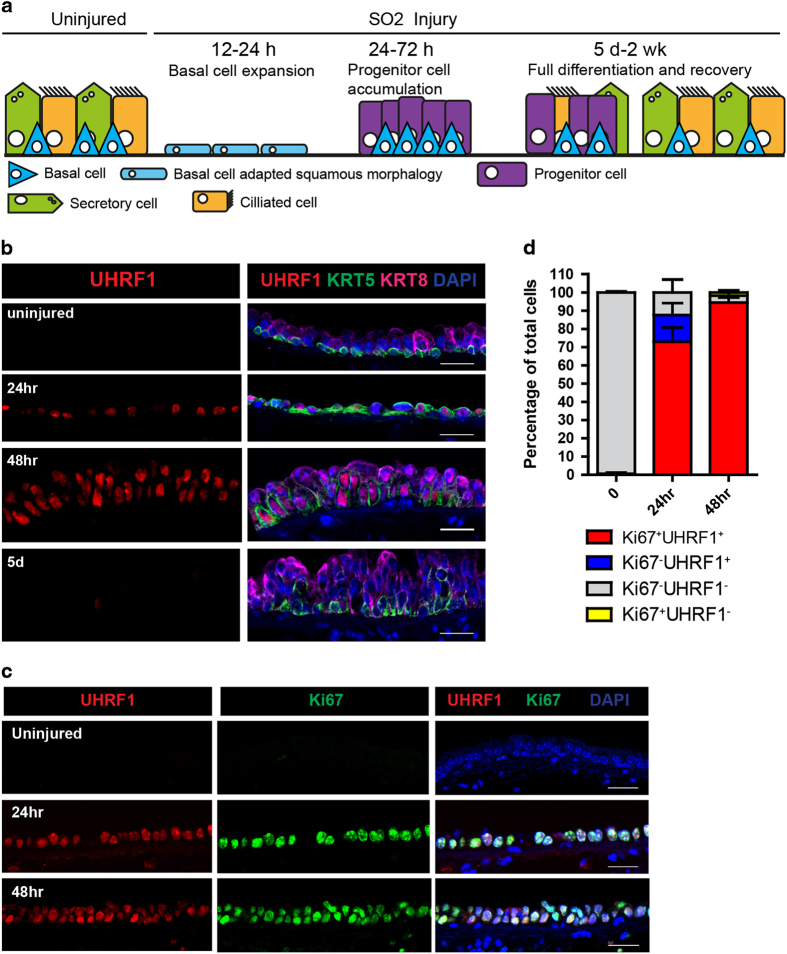
UHRF1 expression is tightly correlated with cell proliferation during airway epithelium repair. (a) Illustration representing the repair of the mouse tracheal epithelium by basal stem cells after SO2 injury. (b) Confocal images of mouse trachea epithelium collected at steady state, 24 h, 48 h and 5 days after SO2 injury. Longitudinal midline sections stained with antibodies to KRT5 (basal cell marker), KRT8 (differentiated luminal cell and early progenitor cell marker) and UHRF1. Note that UHRF1 expression was undetectable at steady state but was markedly upregulated after injury. (c) Confocal images of mouse trachea epithelium collected at steady state, 24 and 48 h after SO2 injury. Tissue sections were co-stained with UHRF1 and Ki67, a proliferation marker. (d) Quantification of the percentage of Ki67+UHRF1+, Ki67+UHRF1−, Ki67−UHRF1+, Ki67−UHRF1− in total cells in the epithelium. Scale bar: 20 μm.

