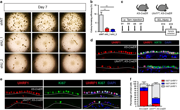
Figure 4.
UHRF1 is required for the proliferation of basal cells in airway epithelium repair and three-dimensional tracheosphere culture. (a) Representative microscopic images of the tracheosphere culture from control (shNT) or UHRF1 knockdown (shU_1 and shU_2) primary HBE cells. Images were taken 7 days after seeding. (b) Quantification of the percentage of colony formation efficiency of each group from a. Data are reported as mean±s.e.m. **P<0.01, P-value was calculated based on three independent experiments. (c) Schematic of loss-of-function (Uhrf1fl/fl;K5-CreER) model. K5-CreER mice and Uhrf1fl/fl;K5-CreER were i.p. injected with four doses of Tmx. One week after the last injection, mice were exposed to SO2, and tracheas were harvested at 24 h after injury. (d) Representative confocal images of tracheas stained with UHRF1 and KRT5 antibodies in control (K5-CreERT) and loss-of-function (Uhrf1fl/fl;K5-CreER) mice. Cells in the epithelium were all KRT5+, indicating the success of the injury. (e) Representative confocal images of tracheas stained for UHRF1 and Ki67 control (K5-CreERT) and loss-of-function (Uhrf1fl/fl;K5-CreER) mice. The percentage of UHRF1+Ki67+ cells significantly decreased in Uhrf1fl/fl;K5-CreER mice. (f) Quantification of the percentage of each indicated cell population. Data are reported as mean±s.e.m. *P<0.05, three mice per group. Scale bar: 20 μm.

