
Fig. 2.
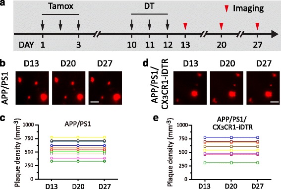
Ablation of microglia by DT administration has no effect on the number of amyloid plaques. a. Timeline of tamoxifen administration, DT administration and in vivo imaging of Congo Red-labeled plaques. Time-lapse imaging was performed 1 day after (D13), 1 week after (D20) and 2 weeks after DT administration (D27). b. Images of the same ROIs in APP/PS1 mice over 1–2 weeks. c. Quantification of plaque density in APP/PS1 mice in each ROI. d. Images of the same ROIs in APP/PS1/CX 3 CR1-iDTR mice over 1–2 weeks. Neither elimination of existing Aβ plaques nor formation of new plaques occurred in APP/PS1 or APP/PS1/CX 3 CR1-iDTR mice. e. Quantification of plaque density in APP/PS1/CX 3 CR1-iDTR mice in each ROI