
Fig. 4.
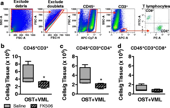
Flow cytometric analysis of T lymphocytes infiltrating skeletal muscle after OST + VML treated with FK506. TA muscles were isolated from OST + VML groups treated with saline or FK506 at 3 days post-injury to evaluate immune cell infiltration by flow cytometry. The middle third region of the TA muscle was collected, digested, and cellular content was isolated. a Events were gated (red polygons) to eliminate debris and doublets, and cells were then gated for CD45+CD3+ T lymphocytes, CD45+CD3+CD8−CD4+ T helper lymphocytes, and CD45+CD3+CD4−CD8+ cytotoxic T lymphocytes. b-d Data for each cell population are normalized per gram of muscle tissue and presented as box (25 to 75 percentile with median line) and whisker (minimum and maximum response) plots; n = 4/group. *p < 0.05