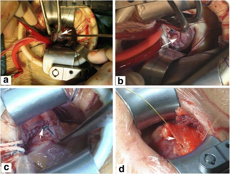
Fig. 3.

Pictures show the closure of subarterial VSD under direct visualization. a After the transpulmonary arteriotomy having been performed, the VSD site was exposed (the arrowhead shows) under direct visualization. b The arrowhead indicates that the subarterial VSD is closing with a bovine pericardial patch. c After the VSD being repaired, pulmonary artery was closed with 5–0 prolene running sutures (the arrowhead shows). d Sequencially, the pericardium was closed loosely with running sutures (the arrowhead shows)
