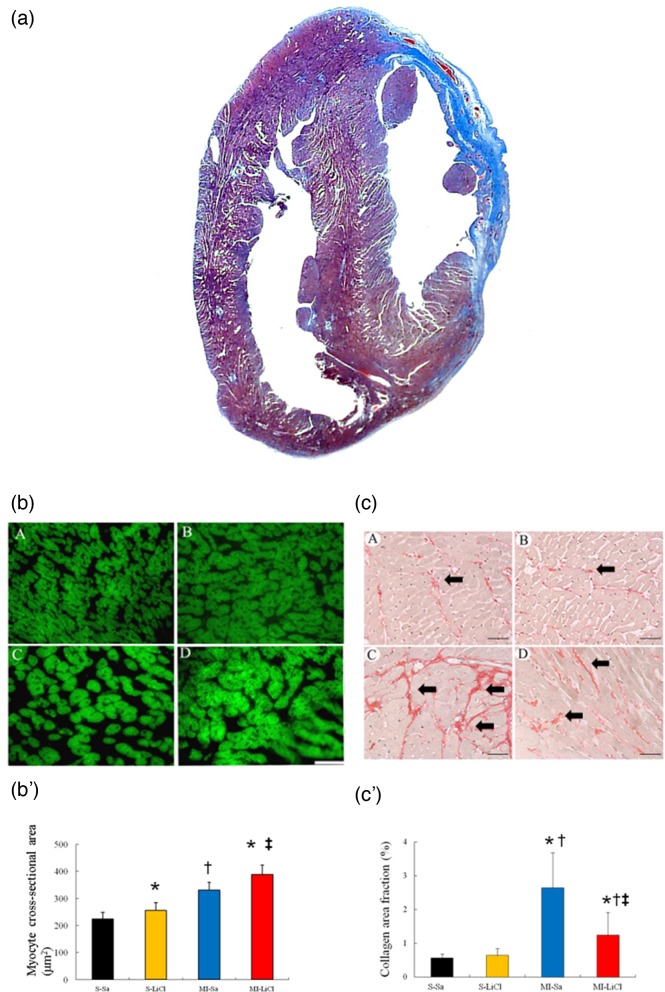
Figure 1. Western analysis of RhoA membrane fraction and cytosolic fraction from the border zone at day 3 after MI.
(a) Representative Masson trichrome-stained section of a vehicle-treated heart at 4 weeks after infarction (blue colour, from 12 to 4 o’clock); Bar =2 mm. (b) Representative cardiomyocytes (magnification 400×) and quantitative analysis of the cardiomyocyte sizes in the remote zone. Staining with FITC-labelled wheat germ haemagglutinin of cross-sectional sections of myocardium; Bar =50 μm. (c) Representative sections from the remote area with Sirius Red staining (red, magnification 400×) at 4 weeks after infarction; Bar =50 μm. (S-Sa), saline-treated sham; (S-LiCl), LiCl-treated sham; (MI-Sa), saline-treated infarcted rat; (MI-LiCl), LiCl-treated infarcted rat. Each column and bar represents mean ± S.D. (n=5–6 per group). *P<0.05 compared with saline-treated sham; †P<0.05 compared with LiCl-treated sham; ‡P<0.05 compared with saline-treated infarcted group

