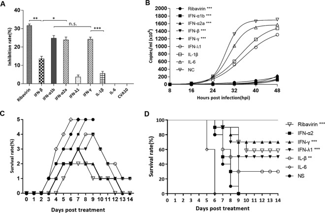
FIG 6.
In vitro antiviral effects of ribavirin, IFN-α1b, IFN-α2a, IFN-β, IFN-γ, IFN-λ1, IL-1β, and IL-6 on CVA10. (A) RD cells were treated with different drugs for 4 h and then infected with CVA10 strain TA151R (MOI = 0.001). After culturing for 24 h, the viral inhibition rate was evaluated using a CCK-8 assay. (B) At 8-h intervals after RD cells were infected with TA151R, 100 μl of culture medium from each experimental and control group was taken out, and viral loads in the supernatant were determined by RT-qPCR. (C and D) Virus loads were expressed as logl0 copies per milliliter, and statistical analysis was performed using one-way ANOVA with a Newman-Keuls multiple-comparison test. Clinical symptoms (C) and survival rates (D) were recorded daily after 5-day-old ICR mice (n = 10 per group) were i.m. challenged with lethal doses of TA151R (240 LD50) until 12 dpi. Within 1 h postinoculation, each mouse was i.m. injected with the indicated cytokines. The data shown are expressed as means and SEM and are representative of at least 3 repeated experiments. The Mantel-Cox log rank test was used to compare the survival rates of pups between drug treatment groups and the medium control group. *, P < 0.05; **, P < 0.01; ***, P < 0.001; n.s., not significant.

