FIG 2.
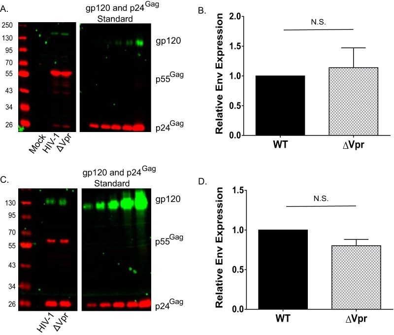
Vpr does not regulate Env expression in infected MDDCs or incorporation of Env into MDDC-derived virions. (A) Western blot analysis of mock-infected, Lai-YU2 (WT)-infected, or Lai-YU2/ΔVpr-infected MDDCs (MOI = 3) for p55Gag and gp120 expression at day 6 postinfection. (B) Quantification of Western blots for p55Gag and gp120 in infected MDDCs, as described above for panel A, from four independent experiments. The gp120 band intensity was quantified and normalized to the p55Gag values from experiments with infected MDDCs derived from 4 donors. Data shown are means (± standard errors of the means). (C) Western blot analysis of p24Gag and gp120 expression in mock-infected, Lai-YU2 (WT)-infected, or Lai-YU2/ΔVpr-infected MDDCs (MOI = 5). MDDC culture supernatants were harvested at days 3, 6, and 9 postinfection; pooled; and concentrated over a 20% sucrose cushion, and virus pellets were lysed for Western blot analysis. (D) Quantification of data from Western blot analysis of MDDC-derived virions from three independent donors. The band intensity for gp120 was quantified and normalized to the p24Gag band intensity. Data shown are means ± (standard errors of the means). Significance was calculated by using a one-sample t test (N.S., not significant [P > 0.05]).

