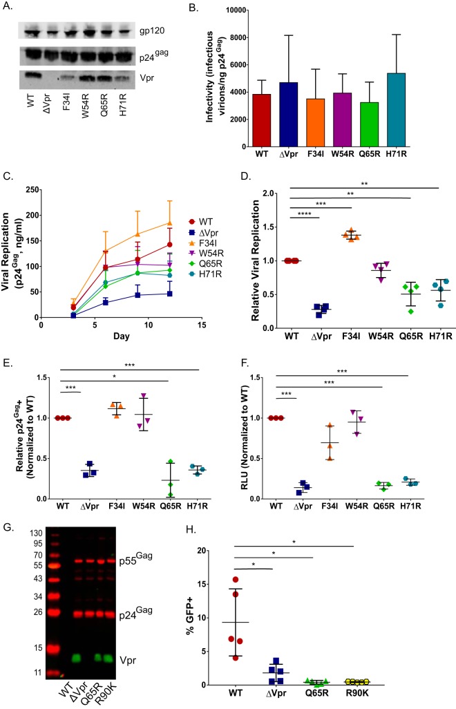
FIG 6.
Vpr mutants deficient for interaction with the DCAF1/DDB1/E3 ubiquitin ligase and inducing G2 cell cycle arrest are attenuated in a single cycle of replication in MDDCs. (A) Representative Western blot analysis of HEK293T-derived Lai-YU2 (WT) and the indicated Vpr mutant viruses used for MDDC infections. Blots were probed with anti-p24Gag, anti-Vpr, and anti-gp120 antibodies. (B) Infectivity of Lai-YU2 and the corresponding Vpr mutants in TZM-bl cells, reported as the number of infectious units (blue cells) per nanogram of p24Gag equivalent, and results are the means (± standard errors of the means) of data from three independent viral preparations. (C) Viral growth curves for four independent infections of MDDCs with Lai-YU2 and the indicated Vpr mutants in DCs. Viral growth was determined by analyzing p24Gag release into cell culture supernatants at days 3, 6, 9, and 12 postinfection by an ELISA. (D) Areas under the curve compiled for four independent MDDC infections represented in panel C, normalized to the value for WT virus infection, which was set to 1 (means ± standard errors of the means). (E) Percentage of p24Gag-positive MDDCs at day 3 postinfection as measured by intracellular p24Gag staining and FACS analysis. Cells were treated with indinavir (1 μM) post-virus exposure to prevent viral spread. The data were normalized to the value for WT virus infection, which was set to 1, and depict the means (± standard errors of the means) of results from three independent infections of MDDCs from three donors. (F) MDDCs infected with 40 ng p24Gag equivalents of Lai-luc Δenv/G (WT or Vpr mutants) were lysed at 3 days postinfection, and viral replication was quantified by measuring luciferase activity in cell lysates. The luciferase activity in Vpr mutant infections was normalized to that of WT virus infections, which was set to 1, and the data shown are the means (± standard errors of the means) for three independent experiments. (G) Western blot analysis of HEK293T-derived Lai-GFP Δenv/G (WT) virus particles or the indicated Vpr mutant virus particles. (H) MDDCs infected with Lai-GFP Δenv/G (WT) or the indicated Vpr mutants (MOI = 3) were harvested at day 3 postinfection and processed for FACS analysis. The data shown are the mean percentages of GFP-positive cells (± standard errors of the means) from five independent experiments with cells derived from five independent donors. Significance was calculated by using paired Student's t test or a one-value t test (comparing normalized data) (*, P < 0.05; **, P < 0.01; ***, P < 0.001; ****, P < 0.0001).

