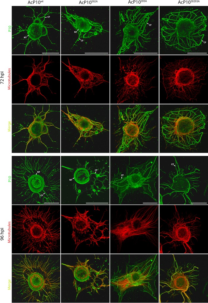
FIG 4.
Analysis of wild-type and mutant P10 structures. TN368 cells were infected with AcP10wt, AcP10S92A, AcP10S93A, or AcP10S9293A and then fixed at 72 and 96 hpi. P10 structures were visualized by using anti-P10- and Alexa Fluor 488 antibodies; microtubules (red) were visualized by using anti-α-tubulin and Alexa Fluor 568 antibodies. P10 and α-tubulin channels were merged to show coalignment. At 72 hpi, cells infected with AcP10wt or AcP10S92A showed both P10 perinuclear tubules (NT) and cytoplasmic filaments (CF). By 96 hpi, the perinuclear tubules had matured, and most cytoplasmic filaments were detached from the central tubule. Cells infected with AcP10S93A or AcP10S9293A lacked perinuclear tubules and displayed rigid and angular cytoplasmic filaments that were not fully detached from the nucleus. Images are representative. Bars, 30 μm.

