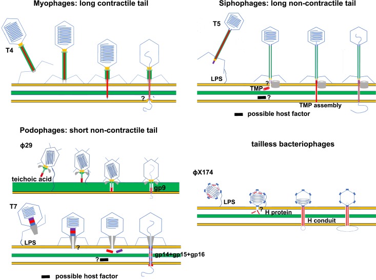
FIG 2.
Schematic diagrams showing host cell entry by prokaryotic viruses. The membrane bilayers are represented by thick yellow lines. The peptidoglycan layers of the bacterial cells are represented by thick green lines or green layers. Membrane-active peptides and proteins are red. The question marks indicate unknown host factors or unclear membrane penetration mechanisms. TMP, tape measure protein; LPS, lipopolysaccharide.

