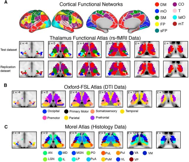
Figure 2.
Cortical functional networks and thalamic atlases. A, Cortical functional networks and thalamic parcellation derived from functional connectivity analyses between the thalamus and each cortical network using rs-fMRI data. Network abbreviations (based on its most predominant anatomical location) are as follows: mO, medial occipital; SM, somato-motor; T, temporal; and latO, lateral occipital. B, Structural connectivity-based segmentation of the thalamus using the Oxford-FSL atlas. Each thalamic subdivision was labeled based on the cortical region it is most structurally connected with. C, Histology-based thalamic parcellation using the Morel atlas. Abbreviations for thalamic nuclei are as follows: MGN, medial geniculate nucleus; PuI, inferior pulvinar nucleus; PuL, lateral pulvinar nucleus; PuA, anterior pulvinar nucleus; Po, posterior nucleus; VM, ventral medial.

