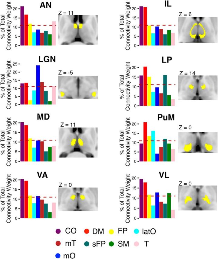
Figure 6.

Distributive pattern of thalamocortical connectivity for thalamic nuclei. Cortical functional networks were most strongly connected with the following thalamic nuclei: AN, LGN, VL, VA, ventral medial (VM), IL, LP, MD, and PuM. Thalamic nuclei (labeled in yellow) are displayed on axial MR images. The bar graphs represent the distribution of connectivity strength between thalamic nuclei and each of the nine cortical functional networks. The dashed line represents the expected proportion of total connectivity if connections were equally distributed across networks.
