Figure 1.

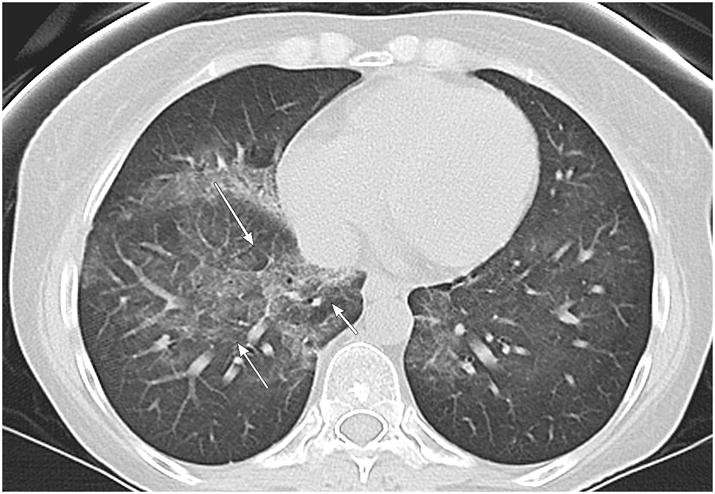
40 year old female with leukemia. Axial CT image demonstrates a diffuse pattern of OP predominately in the right lung lobe with both central and peripheral components (arrows).

40 year old female with leukemia. Axial CT image demonstrates a diffuse pattern of OP predominately in the right lung lobe with both central and peripheral components (arrows).