Figure 3.

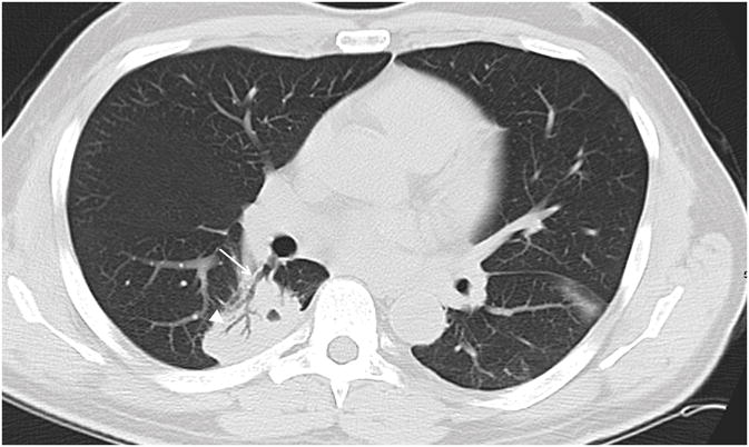
57 year old male patient with leukemia. Axial CT image demonstrates right lower lobe mass like consolidation contacting both central (arrow) and peripheral (arrowhead) bronchi.

57 year old male patient with leukemia. Axial CT image demonstrates right lower lobe mass like consolidation contacting both central (arrow) and peripheral (arrowhead) bronchi.