
Figure 4.
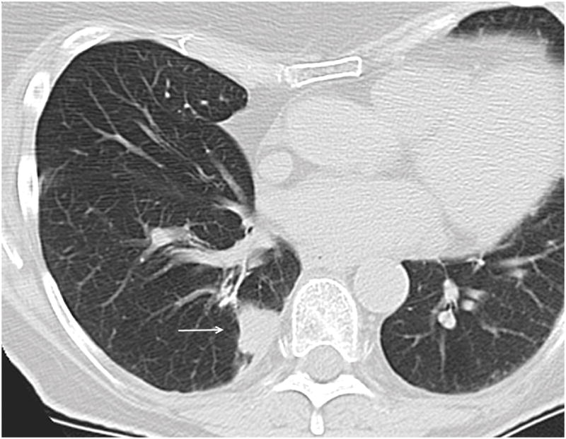
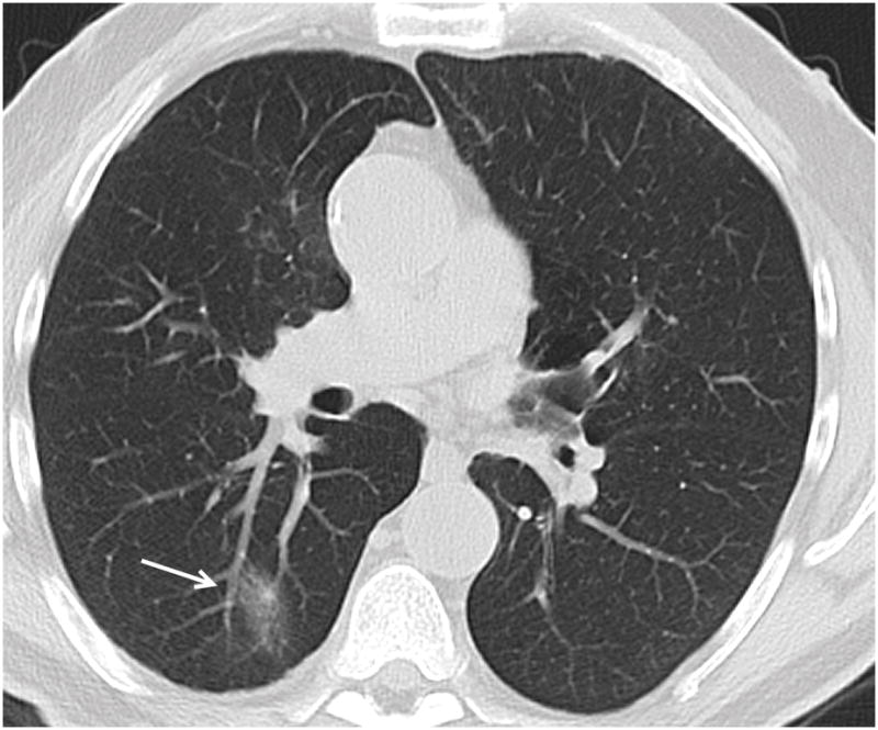
Figure 4a. 74 year old female patient with colon carcinoma. Axial CT image demonstrates OP presenting as a solid lesion in the right lower lobe (arrow).
Figure 4b. 66 year old male patient with head and neck carcinoma. Axial CT image demonstrates OP presenting as a predominantly ground glass nodule in the posterior right lower lobe (arrow).