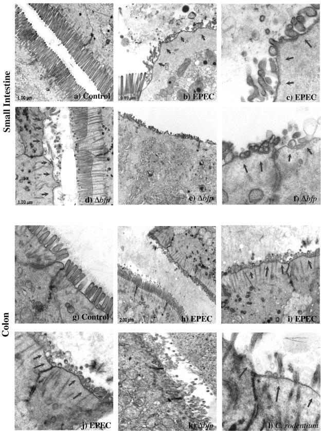
FIG. 3.
EPEC induces microvillous effacement in the intestinal epithelium of infected mice. C57BL/6J mice infected with EPEC were sacrificed after 5 days, and the distal small intestine and proximal colon were examined by TEM. Areas of effaced microvilli and disorganized actin rootlets are highlighted by arrows. The intestinal epithelium of the small intestine (a, ×20,000) and colon (g, ×30,000) in control mice displayed normal microvilli, while that in mice infected with wild-type EPEC (small intestine: b, ×15,000; and c, ×40,000; colon: h, ×1,200; i, ×10,000; and j, ×30,000) or the Δbfp mutant (small intestine: d, ×80,000; e, ×20,000; and f, ×80,000; colon: k, ×25,000) showed dramatic effacement of microvilli. Colon tissue infected with C. rodentium (l, ×60,000) was used as a positive control for microvillous effacement. In some areas, normal microvilli can be seen immediately adjacent to microvilli that have been effaced.

