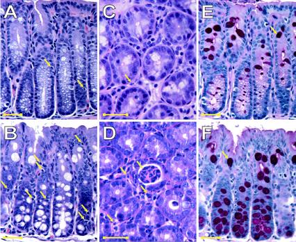
FIG. 5.
EPEC infection induces an inflammatory response in the colon of C57BL/6J mice. The proximal colon of C57BL/6J mice infected with EPEC for 10 days was examined following H&E staining and PAS staining. In control mice, H&E staining of the colon revealed normal mucosal morphology (magnifications: A, ×400; and C, ×1,000). Arrows in panels A and C indicate sparse IEL and lamina propria PMNs in control tissue. However, the colon of EPEC-infected mice showed increases in the numbers of intestinal IELs (B) (magnification, ×400) and lamina propria PMNs (arrows) with crypt abscesses (D) (magnification, ×1,000). PAS staining of goblet cell mucin revealed increased numbers of goblet cells in EPEC-infected colon (F) compared to controls (E) (magnification, ×400). The images shown in panels A and E and B and F were from the same fields. Scale bars in all images represented 25 μm.

