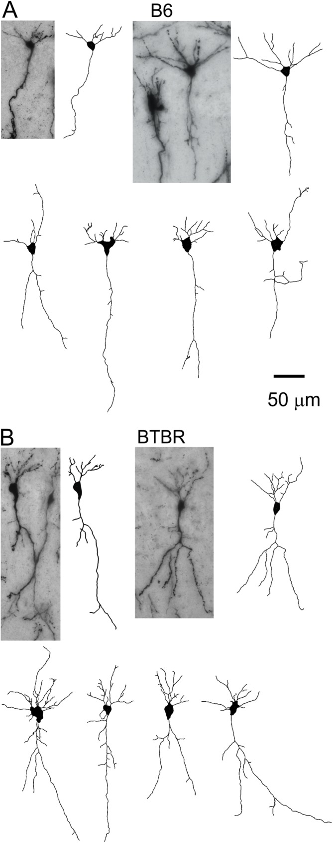
Fig 1. Representative images of Golgi-stained pyramidal neurons in hippocampal CA1 region and the tracings.

(A) First row: two examples of CA1 pyramidal neurons from the B6 animals, and their corresponding tracings. Second row: more examples of tracings from the B6 mice. (B) Similar images of CA1 pyramidal neurons and tracings from the BTBR animals.
