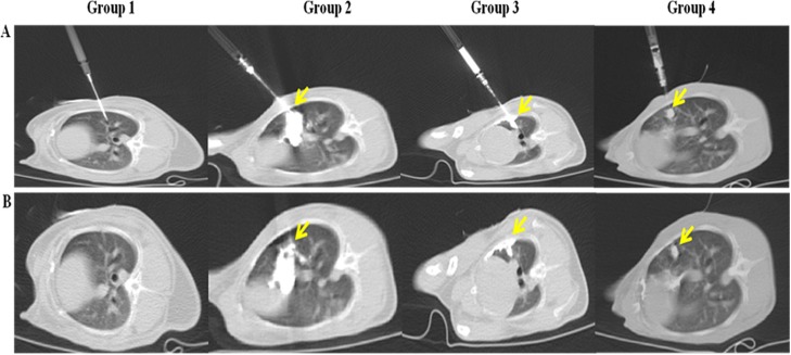
Fig 3. Real-time computed tomography (CT) fluoroscopy-guided percutaneous inoculation of VX2 tumor suspensions.
(A) CT images during inoculation under CT fluoroscopy guidance. (B) CT images after inoculation. The tissue suspensions were not detectable in the images for group 1. In group 2, the tissue suspensions had substantially diffused throughout the lung parenchyma, pleural cavity, and thoracic wall. The cell suspensions in group 3 leaked into the pleural cavity via the inserted needle track. In group 4, the cell suspensions successfully formed globular structures, and there was no leakage. The yellow arrows indicate the suspensions.

