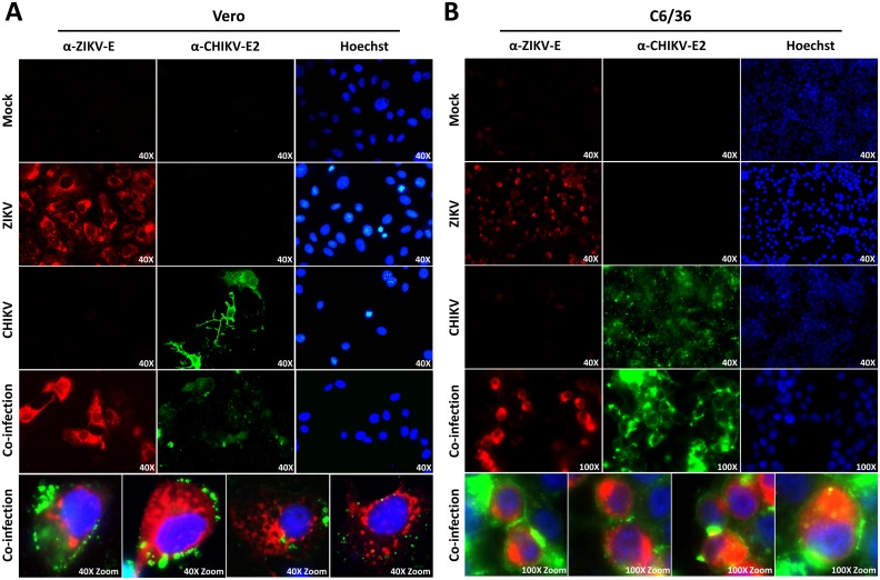
Fig 1. Immunofluorescence of ZIKV and CHIKV single- and co-infections in mammalian and mosquito cells.
(A) Vero and (B) C6/36 cells were infected with ZIKVSUR, CHIKV37997 or both. At 48 hpi the monolayers were fixed, permeabilized, stained with antibodies for ZIKV-E (pan-Flavivirus α-E (4G2)), CHIKV-E2 and Hoechst33258 (details in Materials and methods) and visualized by immunofluorescence. Magnifications are indicated in each picture. Bottom panels indicate zoomed and merged images of co-infected cells.

