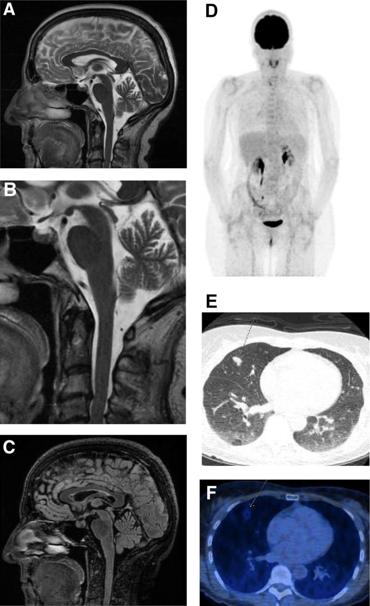
Figure 1.
Staging 32 days before exitus (last staging before exitus) and 21 days before treatment start with pembrolizumab. (A): Magnetic resonance imaging brain: Overview of the brainstem without any signs of inflammation. No swelling or accentuation of the pituitary. The cerebral hemispheres show no focal lesions. (B): Detailed view of brainstem and pituitary gland. (C): Sagittal Fluid‐attenuated Inversion‐Recovery (FLAIR) sequence of the brain with pituitary and brainstem. (D): FDG‐PET/CT overview with several, barely detectable lesions in both lungs, that show minimal metabolic activity. (E): Computed tomography of the lungs with several small nodular infiltrates in the right middle lobe, compatible with metastases or granulomas. Prominent nodular infiltrates in the right middle lobe (arrow). (F): FDG‐PET/CT of the lungs with weak FDG uptake of the above nodular infiltrate.
Abbreviation: FDG‐PET/CT, 18F‐fluorodeoxyglucose (FDG)‐positron emission tomography (PET)/computed tomography (CT).

