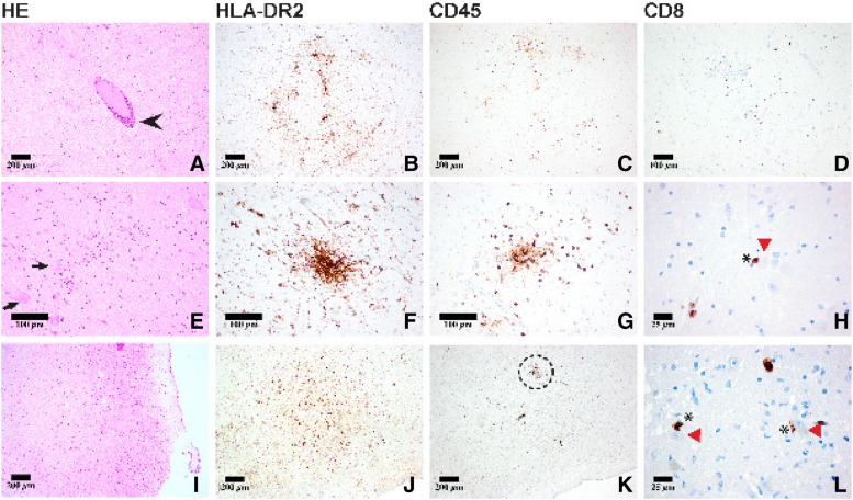
Figure 2.
Neuropathological work‐up of the brain (pons: A–H; I–L: medulla oblongata) showed both perivascular (A, arrowhead) and diffuse (E, arrows: neurons) lymphocytic infiltrates and microglial activation concentrated in the brainstem as highlighted by CD45 and HLA‐DRA2 immunostains, respectively (D, H, I). On occasion, direct proximity to neurons was observed (H, L: asterisk, CD8+ T‐lymphocyte; red arrowhead, neurons in the pons and medulla) suggesting the formation of an “immunological synapse” to important cardiovascular and respiratory neuronal centers in the brainstem (L: close‐up of dashed area seen in K).

