
FIGURE 9:
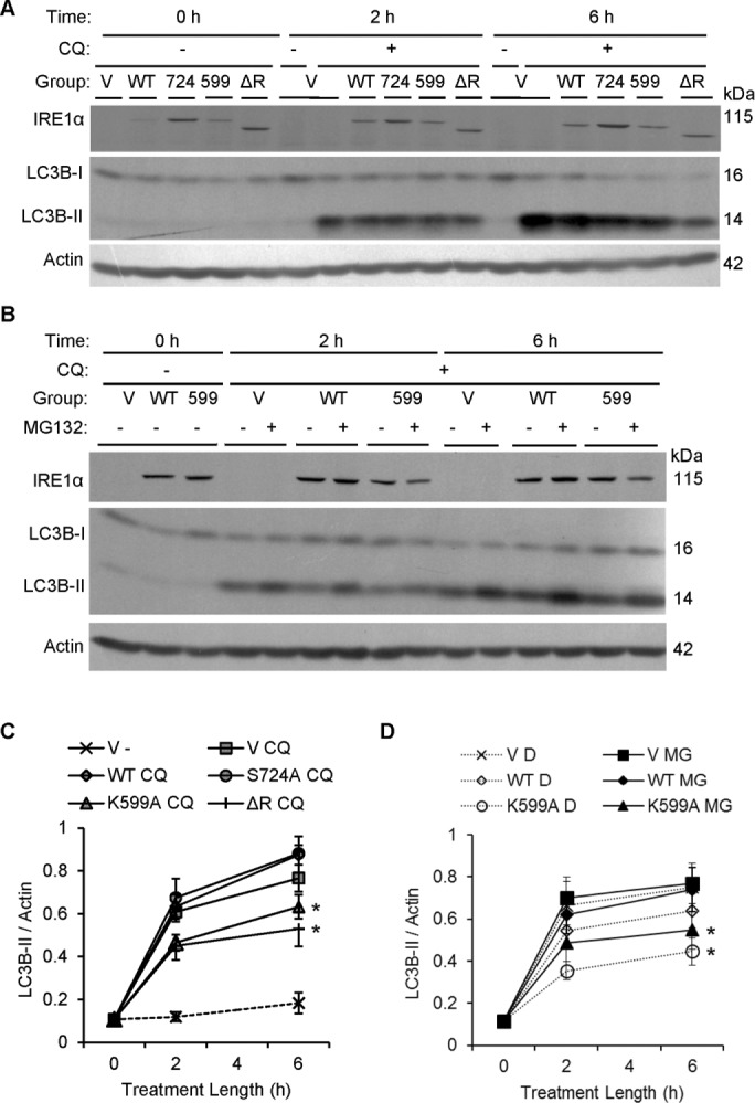
IRE1α facilitates physiological formation of autophagic vacuoles. (A) COS-1 cells transiently transfected with vector (V), FLAG-IRE1α WT, or mutants were treated with or without chloroquine (CQ, 50 μM). Levels of LC3B were measured by immunoblotting after 2 or 6 h. FLAG-IRE1α expression was confirmed using anti-FLAG antibody. (C) Quantification of the rate of LC3B-II accumulation shows that overexpression of IRE1α K599A and ΔR significantly reduces the formation of LC3B-II vs. IRE1α WT. IRE1α ΔR also significantly lowers the rate of LC3B-II formation vs. control (vector), whereas IRE1α K599A tends to lower the rate of formation vs. control (*p = 5.40 × 10⁻⁷ [IRE1α]; p = 1.09 × 10⁻⁹ [CQ]; p < 2.2 × 10⁻¹⁶ [time]; p = 0.0018 [IRE1α × time interaction]; p = 1.07 × 10⁻⁴ [CQ × time interaction]; seven experiments). (B) COS-1 cells transfected with vector (V), IRE1α WT, or K599A were treated with chloroquine (CQ, 50 μM) plus vehicle (DMSO [D]) or MG132 (MG, 25 μM). Overexpression of IRE1α K599A reduced the rate of LC3B-II accumulation independently of MG132 treatment. (D) Quantification of the rate of LC3B-II accumulation over the 6-h chloroquine treatment period shows that overexpression of IRE1α K599A significantly reduced the rate of LC3B-II accumulation vs. control (empty vector) and vs. IRE1α WT. Differences were significant at 2 h and increased further at 6 h (*p = 0.0047 [IRE1α]; p = 1.1 × 10⁻¹⁴ [time]). These effects were independent of MG132. Six experiments.