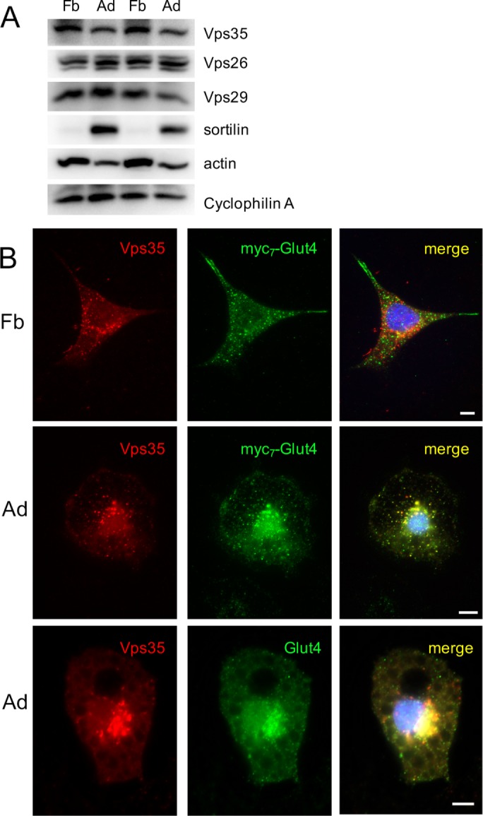
FIGURE 2:

Glut4 is colocalized with Vps35 in differentiated (Ad) but not in undifferentiated (Fb) 3T3-L1 cells. (A) Total lysates (40 µg/lane) of differentiated and undifferentiated 3T3-L1 cells were analyzed by Western blotting. (B) Top and middle, 3T3-L1 cells stably expressing myc7-Glut4 were stained with goat polyclonal antibody against Vps35 and mouse monoclonal anti-myc antibody, followed by Texas red–conjugated donkey anti-goat IgG and FITC-conjugated donkey anti-mouse IgG and analyzed by double immunofluorescence. Bottom, wild-type 3T3-L1 adipocytes were stained with goat polyclonal antibody against Vps35 and rabbit polyclonal antibody against Glut4, followed by Texas red–conjugated donkey anti-goat IgG and FITC-conjugated donkey anti-rabbit IgG.
