FIGURE 1:
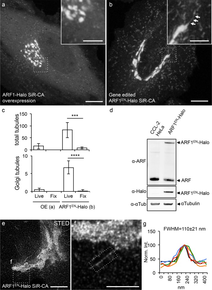
Endogenous tagging of ARF1 highlights Golgi-derived tubular structures. Cells either transiently overexpressing ARF1-Halo (a) or expressing gene-edited ARF1EN-Halo (b) were labeled with SiR-CA and imaged with a confocal microscope. (b) A Golgi-derived tubule is highlighted by arrows. (c) Total numbers of tubules/cell and Golgi-derived tubules/cell were quantified in both cells both live and after fixation with 4% PFA. Result of a two-tailed, unpaired t test. ***p < 0.001, ****p < 0.0001 (n = 10 cells). (d) Gene editing was validated via Western blot using an antibody that recognizes class I ARFs (ARF1 and ARF3) due to the high protein sequence homology. The added amounts of ARF1EN-Halo (∼35%) and unedited ARF (∼70%) in the ARF1EN-Halo cell line match the amount of ARF1 (set to 100%) in CCL-2 HeLa cells. (e, f) ARF1EN-Halo cells were imaged on a custom-built STED setup. (g) The average width (FWHM) of the Golgi tubules was 110 ± 21 nm (n = 20). All STED images were deconvolved; the line profile represents raw image data. All error bars represent SD. Scale bars, 10 μm (a, b), 5 μm (cropped images, a, b), 5 μm (e), 2 μm (f).

