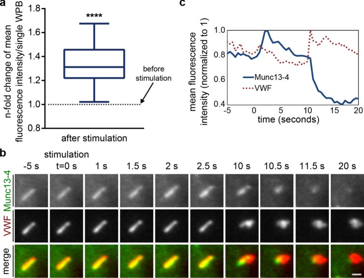
FIGURE 5:
Histamine stimulation induces an additional recruitment of Munc13-4 to WPBs. (a) Munc13-4 fluorescence signals increase on WPBs after histamine stimulation. Cells expressing YFP–Munc13-4 or Munc13-4–mKate together with VWF-RFP or VWF-GFP were stimulated with histamine and imaged by live-cell confocal microscopy. Image stills were thresholded in ImageJ to create ROIs for Munc13-4–positive WPBs in a cell and compare mean fluorescence intensities of all ROIs soon before and soon after stimulation. Mean fluorescence intensity before stimulation was set to 1, and the increase after stimulation was measured as the n-fold change in mean fluorescence intensity for every individual ROIs. Mean n-fold changes from all individual ROIs (a total of 323 WPBs from at least three independent experiments) were tested for statistical significance by one-sample t test (****p ≤ 0.0001). (b) Munc13-4 increases and then disappears at a WPB during exocytosis. HUVECs expressing YFP–Munc13-4 and VWF-RFP were stimulated with 100 µM histamine, and the fusion of individual WPBs with the plasma membrane was recorded by TIRF microscopy. TIRF sections of a single WPB positive for YFP–Munc13-4 and VWF-RFP. The cell was stimulated at t = 0 s, and fusion of this WPB occurred at t = 10 s. See also Supplemental Video Fig5video01. Scale bar, 1 µm. (c) Corresponding mean fluorescence intensities (YFP, RFP) of the WPB shown in b vs. time. The YFP–Munc13-4 signature shows a fluorescence increase on stimulation (t = 0 to 2.5 s) and subsequently a rapid decrease in fluorescence that coincides with the formation of a characteristic VWF fusion spot (t = 10 to 11.5 s).

